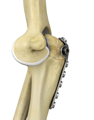
What is Elbow Fracture Reconstruction?
Elbow fracture reconstruction is a surgical procedure employed to repair and restore the appearance and full function of a damaged elbow caused by severe trauma or injury. This may include repairing damaged structures or replacing missing or damaged structures with adjoining skin, muscles, ligaments, tendons, bones, or nerves to restore the appearance and function. This may also include bone fusion (arthrodesis) or replacement of a joint (arthroplasty) to mitigate pain.
Elbow Anatomy
The elbow is a complex hinge joint formed by the articulation of three bones - humerus, radius, and ulna. The upper arm bone or humerus connects the shoulder to the elbow forming the upper portion of the hinge joint. The lower arm or forearm consists of two bones, the radius and the ulna, and connects the wrist to the elbow forming the lower portion of the hinge joint.
What are the Types of Elbow Fractures?
The types of elbow fractures include:
- Radial head and neck fractures: Fractures in the head and neck portion of the radius bone are referred to as radial head and neck fractures.
- Olecranon fractures: These are the most common elbow fractures, occurring at the bony prominence of the ulna.
- Distal humerus fractures: These fractures are common in children and the elderly. Nerves and arteries in the joint may sometimes be injured in these fractures.
Indications for Elbow Fracture Reconstruction
Elbow fracture reconstruction is indicated when conservative measures, such as immobilization of the arm with a cast or splint, physical therapy, rest, injections, or medications fail to resolve the elbow condition.
Diagnosis
Your doctor diagnoses an elbow fracture by performing a physical examination. Other investigations that help diagnose an elbow fracture include:
- X-ray of the elbow is a radiological test carried out to look for abnormalities in bone structures of the joint.
- CT (Computerized tomography) scan of the elbow is done to obtain detailed views of the bone.
- MRI (Magnetic resonance imaging) of the elbow is done to view the bone and surrounding soft tissues.
Preparation for Surgery
Prior to elbow fracture reconstruction, you may have:
- Physical exam to inspect blood circulation and nerves affected by the fracture
- X-ray, CT scan, or MRI scan to assess surrounding structures and broken bone
- Blood tests
- Depending on the type of fracture you have sustained, you may be given a tetanus shot if you are not up to date with your immunizations
- A discussion with an anesthesiologist to determine the type of anesthesia you may undergo
- A discussion with your doctor about the medications and supplements you are taking and if any should be stopped
- A discussion about the need to avoid food and drink past midnight the night prior to your surgery
Elbow Fracture Reconstruction Procedure
Treatment for elbow fractures varies according to the severity and type of the fracture and involves the following:
- External fixation: This method is employed for severe open fractures. During this operation, small cuts are made in the skin and metal pins are inserted through the bones. The pins project out of the skin and are attached to carbon fiber bars outside the skin. The external fixator functions as a frame and aids in holding the elbow in a proper position until a second operation can be performed. It gives damaged skin ample time to heal before surgery to fix the fracture and may decrease the risk of infection.
- Open reduction and internal fixation (ORIF): This is the procedure most commonly used to treat elbow fractures. During this operation, bone segments are first repositioned (reduced) into their normal alignment and then held in place with screws and plates attached to the outside of the bone. Depending on the fracture, your surgeon may have certain considerations during the repair, including:
- Ulnar nerve placement: During the procedure, your surgeon will move the ulnar nerve slightly from its position to make room and to prevent it from being injured during surgery. The nerve is put back into its position at the end of the procedure.
- Bone grafting: If fractured bone has been crushed or lost through the wound, the fracture may require bone grafting to fill the empty bone space. Bone graft may be obtained from a donor (allograft) or from another bone in your own body (autograft) or an artificial material in some cases.
- Osteotomy: In some cases, the surgeon may incise the tip of the elbow (olecranon) to better visualize the bone segments. The incised bone is shifted out of the way during repair of the fracture. After the fracture is secured, the incised olecranon is placed back into its original position and repaired with plates and screws or pins and wire.
- Total elbow replacement (arthroplasty): In cases of severe fractures, the humerus is so badly damaged that it cannot be repaired and needs to be replaced with a metal and plastic implant. During an elbow replacement, after the fragments of bone are removed, an implant is attached to the humerus. Another implant is attached to the ulna (forearm bone), and the two implants are linked to form a hinge.
- Arthrodesis (fusion): In a more active and a younger individual, a severely injured humerus in some cases may be treated with arthrodesis rather than a total elbow replacement. During arthrodesis, your surgeon will apply plates and screws to make the olecranon and humerus grow together or fuse as one bone. Although, the patient will lose the ability to bend the elbow post fusion, but will retain the ability to rotate the hand and regain a strong elbow joint.
Postoperative Care
You may have some pain post procedure and pain medication will be prescribed to keep you comfortable. After surgery, your arm will be placed in a cast or a splint for support and protection. You will need to keep your arm immobile for several weeks with the aid of a sling to allow bone healing. Your doctor will provide instructions on dressings and incision care as well as arm care like application of ice for comfort.
Physical therapy is suggested to prevent arm stiffness, strengthen muscles, and restore range of motion. You will also be advised on a healthy diet and supplements high in vitamin D and calcium to promote bone healing. Your doctor might want you to refrain from certain over-the-counter pain meds as they can hamper bone healing.
Depending on your health condition and the extent of the injury, you may be able to go home the same day with scheduled follow-up appointments for monitoring progress and for stitches or staple removal if necessary. Your doctor may order X-rays to monitor healing throughout your treatment. Most people return to their normal activities within few months.
Risks and Complications
As with any surgery, some of the potential risks and complications of elbow fracture reconstruction may include:
- Bleeding
- Swelling
- Infection
- Pain
- Anesthetic complications
- Damage to nerves and blood vessels
- Hardware irritation
- Nonhealing of fracture
- Broken hardware
- Bone misalignment
- Need for repeat surgery

